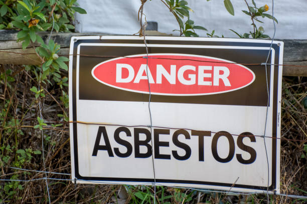
Asbestosis is a chronic lung disease caused by prolonged exposure to asbestos fibers—a once-common mineral used in construction, textiles, mining, and manufacturing. While asbestos use has declined due to health risks, asbestosis remains a significant public health concern, particularly among older workers exposed decades ago in unsafe job sites without proper occupational safety and health protections.
But how is asbestosis diagnosed? Diagnosing this disease requires a multifaceted clinical approach that blends medical history, imaging techniques, lung function tests, and sometimes tissue sampling. Early detection is essential, as asbestosis may progress to more severe conditions such as mesothelioma, asbestos lung cancer, and pleural disease. Let’s break down how physicians and health care providers assess and confirm this serious respiratory system condition.
Medical History and Exposure Assessment
Diagnosis typically begins with a detailed review of the patient’s medical record and occupational history. A physician or pulmonology specialist will ask if the individual worked in the textile, mining, or construction industries, where chrysotile or amphibole asbestos exposure is most likely. Jobs involving floor installation, insulation, manufacturing, or demolition frequently carried high risk.
Clinicians may also explore symptoms such as
- Persistent cough
- Shortness of breath
- Chest tightness or pain
- Fatigue
- Weight loss
- Reduced appetite
- Respiratory infections like influenza
- Wheezing or crackles heard via stethoscope
A thorough medical history ensures that other conditions such as asthma, pneumoconiosis, or tuberculosis are not misdiagnosed in place of asbestosis symptoms.
Physical Examination and Chest Sounds
During a physical examination, doctors listen for crackles—fine, dry sounds in the lower thorax that indicate scarring in the lung parenchyma. They may check for finger clubbing, a sign of reduced oxygen in the bloodstream due to chronic airway obstruction.
Other findings may include:
- Diminished lung volumes
- Evidence of hypoxemia (low blood oxygen)
- Signs of respiratory tract inflammation
- Reduced gas exchange ability
These symptoms guide further investigation into the lung’s internal state.
Radiography: Chest Radiographs and CT Imaging
A chest radiograph (X-ray) is a crucial tool in identifying structural changes in the lungs. In asbestosis, X-rays often show:
- Bilateral, irregular opacities at the lung bases
- Pleural plaques or pleural thickening
- Possible calcification of the pleural tissue
- Atelectasis (lung collapse)
However, chest X-rays alone may lack the sensitivity and specificity needed to confirm a diagnosis. That’s why high-resolution computed tomography (CT) or tomography scans are also employed. CT imaging offers detailed views of fibrosis, scar tissue, and early pleural disease not visible on standard radiographs.
In some cases, CT imaging may also detect pleural effusion—a buildup of fluid between the lungs and thoracic wall—a warning sign of advanced disease or malignancy such as mesothelioma.
Pulmonary Function Testing and Spirometry
To evaluate how much damage has occurred to the lungs, pulmonary function testing (PFT) is conducted. This includes:
- Spirometry—measures how much air the patient can exhale and how fast
- Vital capacity—total air volume the lungs can hold
- Diffusing capacity—checks how well oxygen and carbon dioxide transfer between lungs and blood
Asbestosis often shows a restrictive pattern, meaning the lungs cannot expand fully due to fibrosis and soft tissue stiffening. Reduced airflow and impaired oxygen absorption may indicate advanced disease requiring oxygen therapy or inhaled medication.
Laboratory Tests and Invasive Diagnostics
In cases where imaging and functional testing are inconclusive, doctors may recommend further analysis:
- Bronchoalveolar lavage—samples fluid from the lungs to check for asbestos fiber content and inflammation
- Biopsy or histology—small lung tissue samples are examined under an electron microscope to detect embedded fibers and microscopic pathology.
These procedures help differentiate asbestosis from similar lung diseases and assess progression.
Differential Diagnosis: Ruling Out Other Conditions
Because symptoms can mimic other diseases, diagnosis involves excluding
- Asthma or COPD
- Tuberculosis
- Other pneumoconiosis forms (e.g., silicosis)
- Lung cancer or pleural malignancies
This process requires collaboration among radiologists, pulmonologists, pathologists, and sometimes external organizations like the Agency for Toxic Substances and Disease Registry, American Lung Association, or World Health Organization for guidance and best practices.
Importance of Surveillance and Early Screening
Many people with asbestosis experience a latent period of 10 to 40 years between exposure and symptoms. This makes early screening vital, especially for workers with known contact in Montana, shipyards, or factories where asbestos was widely used.
Annual radiology exams and spirometry can track disease progression and allow for early therapy interventions. Ongoing surveillance improves life expectancy and quality of life.
Management and Therapy
There is currently no cure for asbestosis, but treatment focuses on managing symptoms and preventing complications. This may include:
- Oxygen therapy
- Inhaled steroids or bronchodilators to ease breathing
- Vaccines like the influenza vaccine and pneumococcal vaccine to prevent infections
- Healthy diet to support immune response
- Smoking cessation (if applicable), as tobacco smoke accelerates disease progression
- Pulmonary rehabilitation and nursing support
- Surgical options in advanced cases with pleural effusion or lung collapse
In severe cases, surgery might be needed, although most patients manage symptoms with medication and therapy.
Legal and Support Networks
Patients diagnosed with asbestosis may be entitled to compensation. While our organization is not a law firm, we collaborate with a trusted network of professionals who assist with:
- Lawsuit filing for workplace exposure
- Medical and financial documentation collection
- Referrals to asbestos attorneys and patient advocates
- Resources on the Occupational Safety and Health Administration and United States Environmental Protection Agency standards

Get the Support You Deserve
At the Mesothelioma Asbestos Help Center, we provide access to a nationwide network of medical and legal professionals who understand the unique challenges of asbestos exposure.
Your diagnosis doesn’t have to be the end of the road—it can be the beginning of taking back control of your health, your rights, and your future. Whether you need help understanding your options, seeking second opinions, or navigating compensation resources, our team is here to guide you every step of the way.
Contact us today.

